

Neuroimaging
Neuroimaging in Turkey
Neuroimaging in Turkey includes brain, spine, nerve, and vascular imaging used for diagnosis, treatment planning, follow-up, and surgical preparation. This page explains MRI, CT, CTA, MRA, PET, SPECT, contrast use, implant safety, stroke imaging, seizure evaluation, DICOM files, and cost factors.
What Is Neuroimaging in Turkey?
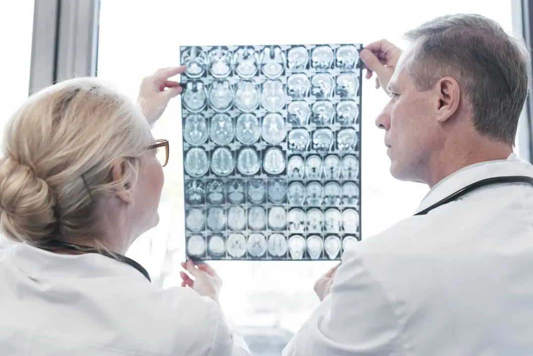
Neuroimaging in Turkey refers to imaging tests that examine the brain, spine, nerves, or related blood vessels. It may include MRI, CT, CTA, MRA, ultrasound, PET, SPECT, or functional imaging in selected cases. Each method answers different clinical questions. CT can be useful in urgent bleeding, trauma, or stroke evaluation. MRI can show detailed brain, spine, soft tissue, inflammation, tumor, or small stroke findings. PET or SPECT can show selected metabolic or blood-flow patterns. The imaging plan should match symptoms, examination findings, urgency, safety factors, and previous records.
Neurosurgery / Neuroimaging
Why Is the Clinical Question More Important Than the Scan Name?
The clinical question is more important because the same symptom can need different imaging pathways. A sudden weakness episode differs from chronic headache, epilepsy, memory change, spine pain, or tumor follow-up. MRI may be preferred for some soft-tissue details. CT may be preferred when speed, bleeding, trauma, or device limits matter. Vascular imaging may be needed when stroke, aneurysm, narrowing, or vessel malformation is suspected. This detail is often missing from competitor pages. Patients should ask what the scan must confirm, exclude, or monitor before scheduling.
Which Symptoms May Require Urgent Neuroimaging?
Sudden neurological symptoms, severe headache, trauma, seizure, or progressive weakness may require urgent neuroimaging.
Face drooping, arm weakness, speech trouble, or sudden vision loss needs urgent care.
A thunderclap headache or headache with confusion needs prompt assessment.
New seizure in an adult may require neurological evaluation and imaging.
Spine pain with weakness or bladder changes should not be delayed.
These signs do not always mean a serious disease is present. They mean timing matters. Emergency assessment should come before travel planning or routine appointment comparison.
When Is Brain MRI Used?
Brain MRI is used when detailed soft-tissue imaging can help evaluate neurological disease. Doctors may request it for tumors, stroke, multiple sclerosis, infection, inflammation, seizures, pituitary disease, memory concerns, or follow-up after treatment. MRI does not use ionizing radiation. However, it still requires safety screening because of the strong magnetic field. Contrast may be added when it improves the clinical answer. A brain MRI protocol can vary by diagnosis. Epilepsy MRI, tumor MRI, pituitary MRI, and stroke MRI are not identical. Correct protocol selection matters as much as scanner availability.
When Is Head CT Used Instead of MRI?
Head CT is often used when fast imaging is needed, especially in urgent settings. CT can help evaluate trauma, bleeding, skull injury, acute stroke pathways, hydrocephalus, and some severe headaches. It is usually faster and less sensitive to patient movement than MRI. CT uses ionizing radiation, so the medical reason should be clear. CT may also involve iodinated contrast in selected cases. MRI may show some brain details better, but CT can be more practical in emergencies. The choice should reflect urgency, suspected diagnosis, safety factors, and available clinical information.
How Do CTA, MRA, and Perfusion Imaging Help?
CTA, MRA, and perfusion imaging help evaluate blood vessels and blood flow in selected neurological conditions.
CTA can show vessel blockage, narrowing, aneurysm, or vascular malformation.
MRA can image many vessels without ionizing radiation.
Perfusion imaging can assess blood-flow patterns in selected stroke pathways.
Vascular imaging may guide urgent treatment or surgical planning discussions.
These tools do not replace clinical judgment. Their value depends on symptom timing, neurological findings, treatment eligibility, contrast suitability, and local protocol.
How Is Neuroimaging Used for Stroke Evaluation?
Stroke neuroimaging is used to distinguish bleeding, blocked vessels, tissue injury, and treatment-relevant patterns. In sudden stroke symptoms, timing is critical. CT may rapidly check for bleeding. CTA may show large vessel blockage. MRI can detect smaller or early ischemic changes in selected settings. Perfusion imaging may help evaluate tissue at risk in defined cases. The scan choice depends on onset time, symptoms, hospital pathway, kidney function, contrast suitability, and treatment options. Patients should not travel for non-urgent imaging during active stroke symptoms. Emergency care should be sought immediately.
How Is Neuroimaging Used for Seizures?
Neuroimaging can help identify structural causes when seizures are new, focal, or clinically concerning. MRI may show tumors, scars, malformations, stroke changes, infection, or inflammation. CT may be used urgently after a first seizure, injury, or altered consciousness. PET or SPECT may be discussed in selected epilepsy surgery evaluations. EEG remains important because it records electrical activity, not anatomy. This difference is often underexplained online. A normal MRI does not exclude epilepsy. An abnormal MRI does not replace neurological assessment. Doctors interpret imaging with seizure description, EEG, labs, medicines, and medical history.
What Are Advanced MRI Techniques and Their Limits?
Advanced MRI techniques can add planning information, but they do not guarantee diagnosis or treatment success. Functional MRI may map selected language or movement areas before brain surgery. Diffusion tensor imaging may estimate white matter tract pathways. MR spectroscopy may assess chemical patterns in selected lesions. Perfusion MRI may help evaluate blood-flow features. These techniques can support tumor, epilepsy, stroke, or surgical planning discussions. They also have limitations from motion, artifacts, analysis methods, and interpretation differences. This is a key missing detail. Advanced imaging should answer a defined clinical question.
Why Do Contrast, Kidney Function, and Pregnancy Status Matter?
Contrast, kidney function, and pregnancy status matter because they can change scan selection and preparation. MRI contrast usually involves gadolinium. CT contrast usually involves iodinated material. Kidney disease can affect contrast decisions. Previous contrast reactions should be reported before the appointment. Possible pregnancy should be discussed before CT or contrast-enhanced studies. Breastfeeding guidance can differ by contrast type and local protocol. Patients should not request contrast because it sounds more complete. Contrast should be used when it adds clinical value. The radiology team should match contrast choice with patient risk.
Why Is Implant and Device Screening Essential?
Implant and device screening is essential because MRI uses a powerful magnetic field. Pacemakers, cochlear implants, aneurysm clips, stimulators, pumps, shrapnel, or metal fragments may need detailed review. Many modern devices are MRI-conditional, but the model and conditions matter. Unknown device details can delay or change the scan. CT may be used when MRI is unsafe or impractical. Patients should bring implant cards, operation notes, or device documentation when available. This point is often missing from tourism pages. Safety screening protects the patient and helps avoid unusable images.
What Records Should International Patients Bring?
International patients should bring complete records so neuroimaging answers the correct clinical question. Useful records include previous MRI, CT, PET, angiography, EEG, surgery notes, pathology, laboratory results, discharge summaries, medicines, and implant documents. DICOM image files are more useful than screenshots or printed images. Prior scans help show whether a finding is new, stable, or changing. Translated summaries can reduce delays when records use another language. Patients should describe symptoms clearly, including timing, side, triggers, weakness, seizures, headaches, and treatment history. Complete records reduce repeated scans and unclear recommendations.
How Should Neuroimaging Results Be Interpreted?
Neuroimaging results should be interpreted by connecting images with symptoms, examination, and prior records. A radiology report may describe lesions, swelling, bleeding, infarct, enhancement, narrowing, degeneration, or comparison findings. Not every abnormality explains symptoms. Age-related changes, incidental findings, old scars, benign cysts, or artifacts can appear. Some urgent diseases may also be subtle early. The referring doctor should relate the report to neurological findings and treatment goals. Further imaging, blood tests, EEG, biopsy, or specialist review may be needed. Patients should avoid making decisions from isolated report wording.
Which Turkey-Specific Health Tourism Rules Matter?
Turkey-specific health tourism rules require information to stay factual, transparent, and non-misleading. Health service promotion should not create demand through guarantees, superiority claims, pressure, or unsupported outcome statements. International health tourism services also involve authorization rules for healthcare facilities and intermediary organizations. Patients may ask whether the provider is authorized for international health tourism. They should request clear information about scan scope, contrast policy, interpreter support, privacy, DICOM transfer, report language, and follow-up communication. This page avoids price promises, comparative savings language, success claims, and unsupported technology superiority statements.
What Should Patients Know About Neuroimaging in Turkey Cost 2026?
Neuroimaging in Turkey Cost 2026 can vary by patient needs, scan type, protocol, contrast use, and reporting requirements. A responsible cost explanation should not use cheapness claims, savings comparisons, or pressure language. The final amount may depend on MRI, CT, CTA, MRA, PET, SPECT, anesthesia needs, urgent reporting, contrast, kidney testing, DICOM transfer, translation, and specialist review. Patients should request written, patient-specific information before travel or booking. Prices should not be presented as a reason to undergo imaging. The clinical indication, safety screening, and correct protocol should come first.
References
NINDS — Neurological Diagnostic Tests and Procedures: https://www.ninds.nih.gov/health-information/disorders/neurological-diagnostic-tests-and-procedures
NINDS — Assess and Treat Stroke: https://www.ninds.nih.gov/health-information/stroke/assess-and-treat
NINDS — Epilepsy and Seizures: https://www.ninds.nih.gov/health-information/disorders/epilepsy-and-seizures
RadiologyInfo — Brain MRI: https://www.radiologyinfo.org/en/info/mri-brain
RadiologyInfo — Head CT: https://www.radiologyinfo.org/en/info/headct
RadiologyInfo — MRI Safety: https://www.radiologyinfo.org/en/info/safety-mr
RadiologyInfo — Contrast Material Safety: https://www.radiologyinfo.org/en/info/safety-contrast
ACR Appropriateness Criteria — Headache: https://acsearch.acr.org/docs/69482/Narrative
ACR — Manual on MR Safety: https://www.acr.org/clinical-resources/radiology-safety/mr-safety
National Cancer Institute — Cancer Imaging Basics: https://dctd.cancer.gov/research/research-areas/imaging/basics
T.C. Sağlık Bakanlığı — Regulation on International Health Tourism and Tourist Health: https://adiyamaneah.saglik.gov.tr/EN-419655/regulation-on-international-health-tourism-and-tourist-health.html
T.C. Sağlık Bakanlığı — Health Tourism Regulations: https://shgmturizmdb.saglik.gov.tr/EN-107627/regulations.html
T.C. Sağlık Bakanlığı — Sağlık Hizmetlerinde Tanıtım ve Bilgilendirme Faaliyetleri Hakkında Yönetmelik: https://antalyaism.saglik.gov.tr/TR-366500/saglik-hizmetlerinde-tanitim-ve-bilgilendirme--faaliyetleri-hakkinda-yonetmelik.html